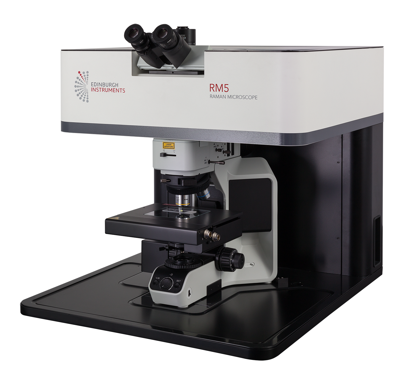
Raman Spectroscopy
Raman Spectroscopy is a non-destructive technique that is used for the identification and quantification of chemical composition. The technique was named after physicist C. V. Raman, Nobel Prize winner in 1930 for contributions to spectroscopy.
A Raman spectrometer radiates a monochromatic light source (a laser beam) into a sample, collecting the scattered light. The majority of scattered rays are in the same frequency as the incoming laser beam, a phenomenon known as elastic or Rayleigh scattering. A very small proportion of the scattered light shifts its energy level and thus frequency, due to the interaction of the electromagnetic waves of the laser beam and vibrational movements of the molecules in the sample. This is called Raman or inelastic scattering.
After plotting this Raman scattering versus frequency, the resulting Raman spectrum is used to calculate a so-called ‘fingerprint’, which is unique for a given compound. The user can then learn the chemical composition and features of the sample by searching the fingerprint against a database or library of known components. Raman spectra reveal vibrational modes of the sample’s chemical components and are therefore an ideal spectroscopic tool for chemical identification.
Combined with an optical microscope, Raman spectroscopy is more sensitive, less affected by unwanted background interactions, and offers the spatial resolution required for chemical mapping.
For further information or to discuss your requirements, please contact us.

Spectral School
Raman spectroscopy an analytical technique where scattered light is used to measure the vibrational energy modes of a sample. Find out more about the theory behind Raman spectroscopy in this article.
How to choose your lasers for Raman spectroscopy
Laser choice is critical for obtaining high quality Raman spectra. This article details the positives and negatives of the three laser regions used in Raman spectroscopy: UV, visible, and near-infrared.
Spectral resolution in Raman spectroscopy
In Raman spectroscopy spectral resolution refers to the ability of the spectrometer to separate neighbouring peaks. This article covers the main factors that affect spectral resolution and the pros and cons of changing these to achieve higher resolution.
What is resonance Raman spectroscopy?
In this article find out about how to gain enhancement in your Raman spectra using resonance Raman, and what applications this technique is most advantageous for.
Surfaced Enhanced Raman Scattering (SERS)
Surface enhanced Raman Scattering (SERS) is a surface sensitive enhancement technique used to obtain higher Raman intensities. This article explores the two mechanisms that occur during SERS enhancement, what substrates can be used, and the combination of resonance Raman with SERS for even greater enhancements.
What is a confocal Raman microscope?
Edinburgh Instruments Raman Microscopes Combe the spectral information from Raman spectroscopy with the spatial filtering of a confocal optical microscope for high-resolution chemical imaging of samples, find out more in this article.
Spatial resolution in Raman spectroscopy
Achieving high spatial resolution in Raman microscopy is crucial for high quality Raman maps. This article details the main factors contributing to lateral and axial resolution.
Role of the pinhole in a confocal microscope
The pinhole is the defining feature of a Confocal Raman Microscope, providing major advantages in spatial resolution and imaging contrast over a conventional optical microscope. In this article read about the role of the pinhole in a Confocal Raman Microscope.
Laser spot size in a microscope
The spot size that a laser can be focussed down to in Raman or fluorescence microscopy is an important parameter that depends on the wavelength of the laser and the properties of the microscope’s objective lens. Find out more in this short article.
The Rayleigh Criterion for Microscope Resolution
The lateral (X-Y) resolution of fluorescence and Raman microscopes is frequently calculated using the famous Rayleigh Criterion for resolution, 0.61λ/NA, but where does this resolution limit arise from and how does it relate to the other resolution limits encountered in microscopy? Find out the answer in this article.
Grating Selection for Raman Spectroscopy
What is the best grating for your Raman spectrometer? Read this technical note to find out! The application team investigates a range of excitation wavelengths, and 5 different gratings to provide an insight into grating selection.
Products
Applications
Raman Spectroscopy can be used in the following applications:
|
|
|
|
|
|
|
|
|
|
Read our latest Raman Application Notes:
- 3D Raman Mapping of a Transdermal Patch
-
Confocal Raman and Photoluminescence Microscopy for Forensic Investigations
You can find all of our Application Notes here.


