Optimisation of SERS for Glucose Sensing
Introduction
Raman spectroscopy is a rapid, non-destructive vibrational spectroscopy technique used for the identification and quantification of chemical composition. One drawback of Raman scattering is a very weak effect with only 1 in 106 incident photons being Raman scattered. The useful Raman signals from the sample can therefore be hidden by Rayleigh scattering, background signal, and noise. Surface-Enhanced Raman Scattering (SERS) is a powerful method to enhance the Raman scattering from a sample and increase the sensitivity of Raman spectroscopy.
The SERS effect was first observed by Fleischmann et al. in 1973 when investigating pyridine adsorbed onto an electrochemically roughened silver surface, though they did not correctly identify what was happening to cause the increased Raman signal.1 This led to the realisation that bringing molecules into proximity (e.g. adsorbing) with arrangements of nanoparticles (NPs), or metal surfaces that have nanoscale roughness, caused an enhancement of their Raman scattering intensity disproportional to the concentration of molecules present.
The exact mechanism behind the SERS effect is still debated to this day, two effects are known to contribute to the enhancement: electromagnetic enhancement, and chemical enhancement. During electromagnetic enhancement, plasmon oscillations perpendicular to the metal surface cause enhancement twice: first, of the incident (excitation) light, and second of the Raman scattered light.2 SERS substrates can be designed to optimise the localised surface plasmons, and the areas these occur at are typically referred to as “hotspots”. An illustration of the SERS effect is shown in Figure 1, where the hotspots are at the tips of the surface.
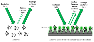
Figure 1. Raman scattering enhancement from a SERS substrate
Chemical enhancement is caused by inter- and intra-molecular charge transfers, and as such the enhancement is greatest of molecules which are adsorbed to the metal surface.3 The enhancement factor for the electromagnetic contribution is on the order of 104, and for the chemical enhancement it is 102, combining to give the 106-fold intensity enhancement of SERS over normal Raman scattering. Typical materials used for SERS substrates are gold and silver because their plasmon frequencies fall within the range of the most common excitation wavelengths used for Raman – visible and NIR.
Since the discovery of SERS, researchers have created many types of SERS substrates, with most falling into two categories – colloidal nanoparticles and nano-patterned surfaces. Focusing on colloids, several of their features affect the enhancement factor achieved. The size of the NPs and their aggregation are studied in this application note for the optimisation of SERS for glucose sensing. Glucose sensing is important for studying diabetes and investigating the effect of foods in blood glucose levels for both healthy and diabetic people.
Materials and methods
To optimise and measure the glucose sensing performance of Au NPs an RM5 Raman Microscope equipped with a 785 nm laser was used. Gold nanoparticles purchased from Sigma Aldrich were used to provide the enhancement.

Figure 2. RM5 Raman Microscope.
Results and Discussion
Nanoparticle diameter
To investigate the relationship between NP diameter and excitation wavelength (785 nm), gold NPs (AuNPs) of a range of diameters (20 nm, 30 nm, 40 nm, 50 nm, 60 nm, 80 nm) were functionalised with 4-nitrothiophenol (4-NTP). Two types of solutions were then made up for each AuNP diameter; solutions where the total number of NPs was held constant, and solutions where the total surface area of the AuNPs was held constant to rule out their effects on increasing the enhancement factor measured. Figure 3A and 3B show the Raman spectra of the 4-NTP band with increasing NP diameter for constant total surface area and constant number of NPs respectively.
For the determination of optimal NP size, the intensity of the 4-NTP peak at 1350 cm-1 was plotted against NP diameter in Figure 3C. This shows that as AuNP diameter increases and approaches the optimum size for the 785 nm excitation wavelength, the enhancement factor also increases.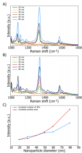
Figure 3. A) Raman spectra highlighting 4-NTP peak with constant area of AuNPs B) Raman spectra highlighting 4-NTP peak with constant number of AuNPs C) Intensity of 4-NTP peak with increasing AuNP diameter.
Aggregation effects on enhancement
After determining the size of the NPs effect on the enhancement factor, aggregation was investigated. An aqueous solution was made containing 1,2(4-pyridyl)ethylene (the analyte), 40 nm AuNPs, and an aggregating agent. The Raman spectra were acquired using a 785 nm laser and studied over a period of time. The software operating the RM5, Ramacle, allows for a kinetic series of measurements to be set up and ran automatically without any further user input. Here, spectra were taken after 0s, 20s, 40s, 160s, and 120s.
Raman spectra in Figure 4A show the increased enhancement seen with NP aggregation over time. The intensity of the peak at approximately 1617 cm-1 was measured and plotted against time, Figure 4B. The data shows that increasing time increases aggregation of the NPs into clusters. This creates hotspots which enhance the Raman scattering signal from the analyte.
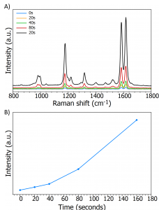
Figure 4. A) 1,2(4-pyridyl)ethylene spectra over time B) Intensity of 1,2(4-pyridyl)ethylene 1617cm-1 peak over time as AuNPs aggregate.
Glucose sensing
A nanosensor can be created by functionalising NPs with molecules that cause the NPs to aggregate in the presence of the desired analyte. This can be used for glucose sensing by functionalising the NPs with mercaptophenylboronic acid (MPBA). This molecule contains a thiol for attachment to the AuNPs, and a boronic acid functional group which can bind to glucose. The binding ratio of MPBA/glucose is 2:1, which brings the nanosensors into proximity with each other, enhancing the Raman signal proportionally to the concentration of glucose present. Spectra were collected using a 785 nm excitation from solutions containing MPBA-AuNPs, glucose of varying concentrations (0.1 – 100 mM), and a buffer of pH 9 (a suitable pH value for MPBA/glucose binding).
The Raman spectra in Figure 5 show the relationship between the intensity of the MPBA peak at approximately 1066 cm-1 and increasing glucose. The peak gains intensity as the glucose concentration rises, this can be used to provide a calibrations plot where the glucose concentration of an unknown sample can be measured.
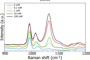
Figure 5. Raman spectra highlighting the MPBA peak intensity at different glucose concentrations.
Conclusion
Since its observation and subsequent discovery over 40 years ago, SERS has developed into its own thriving field covering fundamental and applied uses across chemistry, biology and physics. The enhancement factor provided by SERS substrates allow a Raman spectroscopist to acquire signals from samples that previously scattered too weakly or were in too low a concentration to be seen, and to create nanosensors based on the SERS effect. In this application note we have demonstrated how the RM5 Raman Microscope can be used for the optimisation of physical properties of SERS substrates to increase the enhancement factor and subsequently be used as nanosensors.
References


