Genetically Encoded RNA Sensors for Molecular Imaging in Living Cells
Key Points
- Molecular imaging in live cells is used to monitor cellular processes and signal transduction.
- Genetically encoded RNA sensors, developed by researchers from Hunan University, enabled ratiometric and multiplexed imaging of small molecules in live cells.
- These sensors use aptazyme for recognition and light-up RNA aptamers as signal reporters, allowing versatile imaging of diverse molecules in living cells.
- The stability properties of the sensors were studied via fluorescence analysis using an Edinburgh Instruments FS5 Spectrofluorometer.
- The findings were recently published in Analytical Chemistry.
Introduction
Molecular imaging of small molecules in live cells is essential for monitoring and interpreting their biological processes, such as imaging their cellular metabolites and signal transduction. Genetically encoded fluorescent protein-based biosensors have been developed and used to image various signalling molecules and cellular processes in live cells. However, imaging small molecules remains challenging due to the limited availability of protein domains for small-cell recognition. In addition, fluorescent protein-based sensors suffer from limited dynamic range due to their conformational change upon small-molecule binding.
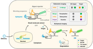
Researchers from Hunan University developed genetically encoded RNA sensors with an innovative modular design that enables ratiometric and multiplexed imaging of small molecules in live cells (Figure 1). These sensors consist of light-up RNA aptamers (such as Broccoli, Mango, and Steroid Receptor RNA Activator Protein 2 or SRB2), which are single-stranded RNA molecules that can bind to specific target molecules with high affinity and specificity. The genetically encoded RNA sensors in live cells demonstrated specific and sensitive responses to small molecules like theophylline, tetracycline, and guanine.

Figure 1: Schematic illustration of the genetically encoded RNA sensors. Adapted with permission from “Genetically Encoded RNA Sensors for Ratiometric and Multiplexed Imaging of Small Molecules in Living Cells”, Ze Fan, Cai-Xia Dou, Li-Juan Tang, Fenglin Wang, and Jian-Hui Jiang, Analytical Chemistry 2023 95 (38), 14455-14464, DOI: 10.1021/acs.analchem.3c03027. Copyright 2024 American Chemical Society.
The researchers used an Edinburgh Instruments FS5 Spectrofluorometer to measure the fluorescence of the sensors. They studied the sensor stability and concentration-dependent response, showing that the fluorescence signals dynamically increased with increasing concentrations of the target molecules and reached a maximum at specific concentrations. This feature indicates the sensors‘ suitability for quantitative molecular imaging and detection of small molecules in a dose-dependent manner.
Experimental Setup
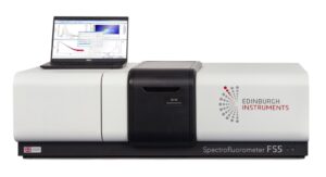
The Edinburgh Instruments FS5 Spectrofluorometer (Figure 2) used in this work was equipped with a 150 W Xenon lamp and a visible PMT-900 photodetector. The samples were held using the SC-05 standard cuvette holder.

Figure 2: Edinburgh Instruments FS5 Spectrofluorometer.
In Vitro Fluorescence with FS5 Spectrofluorometer
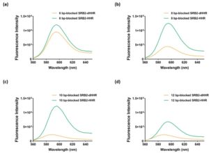
The genetically encoded sensor consisted of an aptazyme sensitive to small molecules, a light-up RNA aptamer as a signal reporter, and a blocking sequence that modulates the sensor’s function by controlling the aptamer’s accessibility to its target molecule. As a signal reporter, the researchers chose the SRB2 aptamer that binds sulforhodamine B-dinitroaniline (SR-DN). The blocking sequence was optimised using a hammerhead ribozyme (HHR) and a degenerate HHR (dHHR), which are RNA molecules that possess catalytic activity and are involved in RNA cleavage reactions. The length of the blocking sequence was varied from 6 nucleotides (nt) to 12 nt with a 2 nt step. To determine the optimal length of the blocking sequence, the researchers performed fluorescence analysis of the sensors using an FS5 Spectrofluorometer.

Figure 3: Fluorescence spectra of the SRB2-dHHR/HHR of a) 6 base pair (bp)-blocked, b) 8 bp-blocked, c) 10 bp-blocked, and d) 12 bp-blocked. Adapted with permission from “Genetically Encoded RNA Sensors for Ratiometric and Multiplexed Imaging of Small Molecules in Living Cells”, Ze Fan, Cai-Xia Dou, Li-Juan Tang, Fenglin Wang, and Jian-Hui Jiang, Analytical Chemistry 2023 95 (38), 14455-14464, DOI: 10.1021/acs.analchem.3c03027. Copyright 2024 American Chemical Society.
The FS5’s primary excitation source, a 150 W Xenon lamp, was used at 560 nm to acquire the fluorescence response of the sensors with varied numbers of nt, either with HHR-mediated cleavage (green curves in Figure 3) or dHHR-mediated cleavage (orange curves). The excitation and emission bandwidths were set at 5 nm. The emission range was between 565 and 650 nm. The fluorescence analysis showed that the SRB2 fluorescence gradually decreased as the length of the blocking sequence increased. The SRB2 with a 10 nt blocking sequence (Figure 3c) exhibited the highest signal-to-background ratio after HHR-mediated cleavage. This result allowed the researchers to investigate the 10 nt blocked SRB2 for live cell imaging.
Conclusion
An Edinburgh Instruments FS5 Spectrofluorometer was used to acquire the fluorescence spectra of genetically encoded RNA sensors. The results enabled researchers at Hunan University to facilitate their study of developing such sensors for molecular imaging of small molecules in live cells. The sensors exhibited negligible fluorescence signals in the absence of the target molecules. Upon exposure to the specific target small molecules, the sensors showed a significant increase in fluorescence intensity, indicating their ability to detect and image these compounds selectively.
Full Publication
The results presented in this Research Highlight were published in Analytical Chemistry, and more details are available at https://pubs.acs.org/doi/10.1021/acs.analchem.3c03027.
Thank you for your interest in this resource.
To download , please complete your details.


